Complete Hydatidiform Mole
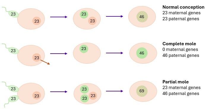
Histologically, a complete hydatidiform mole is characterized by enlarged, edematous, hydropic chorionic villi, often with cistern formation. In addition, there is a marked, usually circumferential, villous trophoblastic proliferation with cytologic atypia.
In very early complete hydatidiform moles, these characteristic histologic features may be less well developed, and trophoblastic proliferation is typically more limited.
As a rule, fetal tissues are absent.Immunohistochemically, p57 expression is absent in the villous stromal cells and the cytotrophoblast.
Partial Hydatidiform Mole
A partial hydatidiform mole typically demonstrates a mixed population of chorionic villi, consisting of small fibrotic villi and enlarged, irregularly shaped villi with variable degrees of edema and hydropic change. Mild to moderate circumferential trophoblastic proliferation is usually present. Cistern formation and trophoblastic pseudoinclusions are common findings.
Fetal blood vessels and nucleated red blood cells are frequently identified.Immunohistochemically, p57 expression is retained in the villous stromal cells and the cytotrophoblast.
The morphological features of a partial hydatidiform mole are not entirely specific and may show considerable overlap with those of an (early) complete hydatidiform mole, hydropic abortion, trisomy syndromes and other chromosomal abnormalities, placental mesenchymal dysplasia, and twin pregnancies with a complete hydatidiform mole coexisting with a normal fetus.
Gestational Choriocarcinoma
Gestational choriocarcinoma is characterized by infiltrative, destructive solid aggregates of mononuclear cytotrophoblast and intermediate trophoblast surrounded by multinucleated syncytiotrophoblast exhibiting marked cytologic atypia and high mitotic activity.
Hemorrhage, necrosis, and lymphovascular invasion are common findings.
Gestational choriocarcinoma may occur in association with an epithelioid trophoblastic tumor and a placental site trophoblastic tumor.
Placental Site Trophoblastic Tumor (PSTT)
Placental site trophoblastic tumor (PSTT) consists of an infiltrative proliferation of implantation-site intermediate (extravillous) trophoblastic cells arranged in nests, cords, and diffuse aggregates infiltrating between the smooth muscle fibers of the myometrium.
The tumor cells are typically large, polygonal, and predominantly mononuclear, with abundant eosinophilic to clear cytoplasm and variable nuclear pleomorphism. Mitotic activity is generally low (1–2 mitoses/mm²).
A characteristic feature is prominent vascular invasion, in which tumor cells replace the vessel wall ("vascular remodeling"), analogous to the physiological process of implantation. Necrosis and hemorrhage may also be present.
Epithelioid Trophoblastic Tumor (ETT)
Epithelioid trophoblastic tumor (ETT) is a neoplasm derived from chorionic-type intermediate trophoblast. Histologically, it typically exhibits a nodular, expansile growth pattern composed of large nests and cords of relatively uniform epithelioid trophoblastic cells with well-defined cell borders.
The tumor cells are mononuclear, medium-sized, and epithelioid, with moderately eosinophilic to clear cytoplasm and a frequently squamoid (carcinoma-like) appearance. Mitotic activity is highly variable.
A characteristic feature is the presence of extracellular eosinophilic hyaline (fibrinoid) material located centrally within the nests or between the tumor cells. Extensive geographic necrosis is frequently observed.