aria.labels.skip_content

Frequently asked questions

What is a gestational trophoblastic disease?


Gestational trophoblastic diseases (GTDs) are a very rare group of conditions that occur when there is an abnormal growth of a fertilized egg inside the uterus. The term “trophoblastic” refers to the trophoblast cells, which surround the fertilized egg. These cells help the embryo attach to the uterine wall and later develop into the placenta

There are two main types of GTD:
 
  • Hydatidiform mole (molar pregnancy): 
    This is the most common form of GTD, accounting for about 80% of all cases. It is a benign condition, meaning it is not cancerous, but it is considered pre-malignant, as it can sometimes develop into a malignant form. Even if it becomes invasive, it remains highly treatable.
    (More information on this type can be found below.)
     
  • Gestational trophoblastic neoplasia (GTN):
    This is a rarer and malignant form of GTD, meaning it is cancerous. However, it is usually completely curable with appropriate treatment and follow-up.
    (More information on this type can be found below.)
Information on the different types of trophoblastic diseases?


A molar pregnancy is part of the spectrum of Gestational Trophoblastic Diseases (GTDs). It is a rare condition, occurring in about 1 in 1,000 pregnancies in Europe. A molar pregnancy develops due to a problem that happens at the time of conception

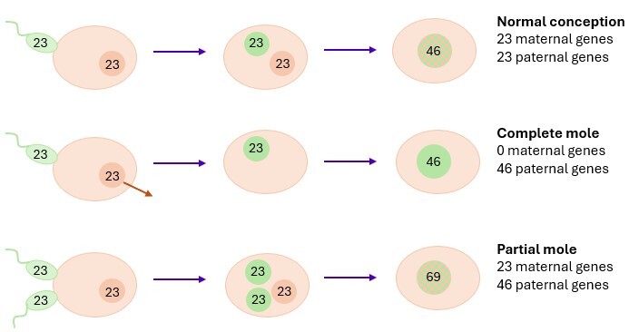
In a normal conception, an egg is fertilized by a single sperm cell. The fertilized egg then begins to divide rapidly and develops into both the placenta and the embryo

In a molar pregnancy, however, there is a genetic imbalance that causes the fertilized egg to grow abnormally. Instead of developing into a normal pregnancy, the tissue grows uncontrollably, forming fluid-filled cysts that look like a cluster of grapes inside the uterus.

 There are two types of molar pregnancies, depending on the kind of genetic problem that occurs at fertilization:
 
  • Complete mole
A complete mole develops when an egg that has lost its maternal DNA is fertilized. This loss of DNA can occur either during the development of the egg in the ovary or at the moment of conception.

Because the egg contains no genetic material from the mother, abnormal fertilization follows one of two possible paths:
 
  • In most cases, a single sperm fertilizes the empty egg and its 23 chromosomes duplicate, forming a full set of 46 chromosomes — all from the father.
  • Less commonly, the empty egg is fertilized by two sperm cells, each contributing 23 chromosomes, also resulting in a 46-chromosome conceptus entirely of paternal origin.

Since all the genetic material comes from the father, the fertilized egg cannot develop into a normal embryo or placenta. Instead, it grows into a mass of rapidly dividing cells, forming clusters of fluid-filled cysts inside the uterus. 

In most cases, removal of the molar tissue is enough to resolve the condition. However:
 
  • Around 15–20% of women may develop a post-molar gestational trophoblastic neoplasia.
  • Of whom, 3% are choriocarcinomas, a rare but malignant form of gestational trophoblastic disease.

(More information about these conditions can be found in the section “What is a GTN?”).

In very rare cases, a twin pregnancy may occur in which one twin is a complete mole and the other is a normal fetus. Unfortunately, these pregnancies often end in miscarriage, with a live birth of the healthy twin in only about 40% of cases
 
  • Partial mole
A partial mole occurs when a single egg is fertilized by two sperm cells at the same time. As a result, the fertilized egg contains 69 chromosomes instead of the normal 46. This genetic imbalance prevents pregnancy from developing normally.

Sometimes, an ultrasound may show an embryo, but it will always have severe abnormalities and will not be viable

In most cases, removal of the molar tissue is enough to resolve the condition. However:
 
  • About 3% of women may develop a post-molar gestational trophoblastic neoplasia.
  • Of whom, 0.5% are choriocarcinoma, a rare but malignant form of gestational trophoblastic disease.

(More information about these conditions can be found in the section “What is a GTN?”).

In very rare cases, a twin pregnancy may occur in which one twin is a partial mole and the other is a normal fetus. Unfortunately, these pregnancies often end in miscarriage, with a live birth of the unaffected twin in only about 40% of cases.


gestational trophoblastic neoplasm (GTN) is part of the spectrum of Gestational Trophoblastic Diseases (GTDs). Several different types of GTNs have been identified, all of which are very rare, occurring in approximately 1 in 40,000 to 1 in 100,000 pregnancies. 

A diagnosis of a GTN can be made biologically, through the monitoring of the hCG (human chorionic gonadotropin) hormone, or histologically, by examining tissue samples under the microscope. 

Although all types of GTN are considered malignant—and therefore cancerous—they are highly curable, with a success rate of around 98% when diagnosis, treatment, and follow-up are performed appropriately in expert teams. 
 
  • Invasive mole (IM)
An invasive mole usually develops from a complete mole, but it can also arise, more rarely, from a partial molar pregnancy. It occurs in about 15–20% of women after a complete mole and in about 3% after a partial mole.

It is called an invasive mole because the molar tissue grows into the muscular wall of the uterus (the myometrium). 
Although it is classified as a malignant form of gestational trophoblastic disease, it rarely spreads outside the uterus

Uterine bleeding is often the main symptom, caused by invasion of the uterine muscle.
The diagnosis is usually made through monitoring of hCG hormone levels (see section 'Diagnosis').

An invasive mole is very well treatable, and chemotherapy or surgery is almost always effective (see section 'Treatment' for more information). 
 
  • Gestational Choriocarcinoma (GCC)
choriocarcinoma is a rare type of gestational trophoblastic neoplasm, occurring in approximately 1 to 9 per 40,000 pregnancies
It usually develops from a molar pregnancy (3% of complete moles and 0.5% of partial moles), but it may also appear after a non-molar miscarriage, an induced abortion, or even a normal pregnancy.

This is an aggressive tumor that grows rapidly and can spread to other organs. Clinical symptoms depend on the extent of the disease:
 
  • Uterine bleeding is common in localized cases.
  • If metastases are present, symptoms usually relate to the affected organs — most often the lungslivervaginakidneys, or central nervous system.

persistently elevated hCG level, combined with compatible symptoms, is key to establishing the diagnosis (see section 'Diagnosis').

Despite its malignant nature, it is highly sensitive to chemotherapy and almost always curable with appropriate treatment  (More information on management can be found in the section 'Treatment') .
 
  • Placental Site Trophoblastic Tumor (PSTT)
Placental Site Trophoblastic Tumors are an even rarer form of Gestational Trophoblastic Neoplasia (GTN), accounting for only about 2% of all cases. They develop from the placental implantation site cells and most often occur after a normal pregnancy. However, they may also arise after a miscarriage, an induced abortion, or following a molar pregnancy.

Unlike other types of GTN, PSTTs are slow-growing tumors, and symptoms may not appear until months or even years after the preceding pregnancy. The average interval between the pregnancy and diagnosis is approximately 3.4 years.

This type of tumor usually remains confined to the uterus, although it can invade the muscular wall (myometrium). The diagnosis is often made following abnormal uterine bleeding or absence of normal menstruation

Levels of hCG are typically lower than in other types of GTN. (More information on diagnosis can be found in the section Diagnosis).

PSTTs are generally treatable, but they are sometimes less responsive to chemotherapy than other GTNs. In such cases, surgical removal of the uterus (hysterectomy) may be required if the disease is confined to the uterus (More details on management are available in the section 'Treatment').
 
  • Epithelioid Trophoblastic Tumor (ETT)
Epithelioid Trophoblastic Tumors are extremely rare forms of Gestational Trophoblastic Neoplasia (GTN). They usually develop after a normal pregnancy, although a small number of cases arise after a molar pregnancy or a spontaneous miscarriage.

Because this is a slow-growing tumor, there is often a long delay — sometimes several years — between the initial pregnancy and the appearance of symptoms.

The diagnosis is often made following episodes of abnormal uterine bleeding or absence of normal menstruation. Levels of hCG are typically only mildly elevated in this type of GTN (More information can be found in the section 'Diagnosis').

ETTs are potentially curable, but they can be less responsive to chemotherapy then other types of GTN. When the disease is confined to the uterus, surgical removal of the uterus (hysterectomy) may be required (Further information on management is available in the section 'Treatment').

molar pregnancy is usually a benign condition. However, in some cases, it can progress to a post-molar neoplasia (GTN), which is a malignant or cancerous form of the disease. This transformation occurs when molar cells are not completely cleared after evacuation and instead continue to grow
For this reason, a molar pregnancy is often described as a precancerous or premalignant condition. 

The exact reasons for this progression are not fully understood, but several risk factors are known to increase the likelihood of developing GTN:
 
  • Maternal age below 20 years or above 40 years
  • Very high pre-evacuation hCG levels (typically >100,000 IU/L)
  • Uterine size larger than expected for gestational age
  • Ovarian theca-lutein cysts
  • Pregnancy complications associated with high hCG levels, such as:
  • Severe nausea and vomiting (hyperemesis gravidarum)
  • High blood pressure or preeclampsia
  • Hyperthyroidism

Although GTNs are malignant diseases, they are highly curable. With appropriate treatment and close follow-up according to clinical guidelines, over 98% of patients are completely cured.
(More information on GTNs can be found in the corresponding section.)

Diagnosis


Many of the symptoms of a molar pregnancy are similar to those of a normal pregnancy — such as morning sickness, vaginal bleeding or abdominal pain. This similarity often makes it difficult to distinguish between the two in the early stages.

The diagnosis is most often made during the first ultrasound scan. The typical ultrasound appearance of a molar pregnancy shows numerous fluid-filled cysts, giving it a “grape-like” or “snowstorm” pattern.
 
  • In a complete mole, a large mass of these cystic structures is seen, with no fetus present.
  • In a partial mole, a fetus and placenta may be visible, but both are abnormal, and the fetus cannot survive.

Women with a molar pregnancy typically have hCG (human chorionic gonadotropin) levels that are much higher than expected for the gestational age. This elevated hormone level explains symptoms such as severe morning sicknesshigh blood pressure, or overactive thyroid function (hyperthyroidism) that may occur in some patients. 

In cases of miscarriage, any tissue collected is routinely examined by a pathologist to determine whether it represents a trophoblastic disease or if another cause of miscarriage can be identified.

In case of ultrasound suspicion of a molar pregnancy, the following steps are recommended:
 
  • Blood tests, including a serum total hCG measurement (the hormone produced in early pregnancy, and the key marker for trophoblastic disease).
  • In some cases, an additional blood test for thyroid function may be required, as high hCG levels can lead to temporary overactivity of the thyroid gland (hyperthyroidism).
  • Physical and gynecological examinations
  • Uterine evacuation by suction under ultrasound guidance
  • Intravenous administration of a uterotonic agent (oxytocin) during suction is encouraged
  • Anti-D prophylaxis must be administered to Rh-negative patients

At this stage, we do not recommend any imaging-based work-up

The diagnosis is established through pathological examination of the curettage specimen. This diagnosis must be confirmed by systematic review by the expert panel of the Belgian Registry of Gestational Trophoblastic Diseases. 

The cornerstone of follow-up in all trophoblastic diseases is monitoring the blood level of hCG (human chorionic gonadotropin), the hormone produced during early pregnancy.  

Although this may feel burdensome at a difficult time, strict adherence to the schedule is essential. It allows your medical team to detect early whether a repeat curettage is needed or whether the disease might be progressing toward a GTN. 
(More information on hCG follow-up schedules can be found in the section 'Follow-up' below.).

An ultrasound check may be performed about 15 days after the initial evacuation to confirm that the uterus is empty. It may show that trophoblastic tissue remains in the uterus. In this situation, a specialist will assess whether a repeat curettage (uterine evacuation) is necessary to remove the remaining tissue.  

Apart from a complete uterine evacuation and hCG monitoring, no other treatment is required for a molar pregnancy unless complications occur.

After a hydatidiform mole diagnosis, serum hCG should be monitored at least once every 2 weeks until normalization (More information in the 'Follow-up' section).  

Gestational Trophoblastic Neoplasia (GTN) is suspected when the hCG levels fail to decline appropriately or begin to rise again during follow-up.

The first essential investigations when a GTN is diagnosed include:
 
  • Physical examination.
  • Ultrasound examination of the uterus, to check for any remaining trophoblastic tissue. Repeat curettage (uterine evacuation) may be considered if residual molar tissue is detected.
  • Gynecological examination, to assess whether the disease has spread to the vagina or pelvis.
  • Blood test to measure hCG (human chorionic gonadotropin)
  • Further imaging will be performed to check whether the disease has spread to other parts of the body. These tests may include:
    • Chest X-ray, to look for spread to the lungs (the most common site of metastasis)
    • CT scan or MRI scan of the abdomen and pelvis
    • MRI scan of the brain

Based on the results of these investigations, your specialist team will determine the most appropriate treatment plan and follow-up schedule for your specific situation.

Because gestational trophoblastic diseases are rare, heterogeneous and complex, challenges are often encountered in their diagnosis, follow-up and treatment.

Accurate diagnosis is essential, as the type of trophoblastic disease — whether a molar pregnancy, invasive molechoriocarcinomaPSTT or ETT — determines the entire management plan

Depending on the specific subtype, the treatment approach can differ completely, ranging from simple monitoring after evacuation to chemotherapy or surgery. Likewise, the recommended waiting period before a future pregnancy varies according to the diagnosis and treatment received. 

It is also important to note that distinguishing between a normal hydropic miscarriage and a molar pregnancy can be challenging. However, this distinction is critical, as it has major implications for follow-up, prognosis and fertility planning

For these reasons, the involvement of specialized reference centers is crucial. Their expertise helps to reduce diagnostic errors and to avoid both under- and over-treatment, ensuring that each patient receives appropriate, individualized care.

Molar pregnancies and GTNs are very rare diseases. Because of this, most gynecologists and pathologists will only encounter a few cases in their entire career. As a result, they may not always have enough experience to accurately diagnose, monitor and treat these conditions. 

To ensure that every patient receives the best possible care, a Belgian Registry for Gestational Trophoblastic Diseases has been created in 2012.

Our mission is twofold:
 
  • To guarantee that all patients receive optimal diagnosis, follow-up, and—when necessary—treatment, specifically tailored to their individual situation.
  • To centralize all cases nationwide, allowing experts to expand medical knowledge and continually improve the diagnosis, follow-up, and treatment of future patients. 

If you agree to be included in the registry, you allow us to:
 
  • Request a sample of the collected tissue, which will be reviewed by an experienced pathologist to confirm or refine your diagnosis.
  • Monitor your hCG levels, to ensure they decrease as expected; if they do not, or if they start to rise again, we will advise your doctor on the next steps.
  • Collect basic background information to help build a profile of all patients and improve our understanding of the disease.
  • Contact you to complete a brief questionnaire that contributes to research and quality improvement.

Participation in the registry is voluntary, and all data are handled confidentially according to the Belgian and European data protection regulations (GDPR).

By agreeing to register your gestational trophoblastic disease, you gain several direct benefits:
 
  • Expert confirmation of your diagnosis:
    Your tissue sample will be reviewed by a specialist pathologist who has experience in gestational trophoblastic diseases, ensuring the most accurate diagnosis possible.
     
  • Specialized medical supervision:
    Your case will be continuously monitored by an expert gynecologist from a reference center, guaranteeing that you receive the most appropriate follow-up and, if needed, treatment tailored to your specific situation.
     
  • A dedicated point of contact:
    You will have direct access to a specialized team at the reference center, where you can ask questions, share concerns, or seek guidance at any stage of your follow-up.

We first recommend that you discuss this with your treating physician. The registration process may simply have been overlooked at the time of your diagnosis. Clarifying this directly helps to maintain a good relationship with your doctor and ensures that everyone involved in your care is informed. 

If, for any reason, your physician declines or omits to register your case, you are strongly encouraged to register yourself. You can easily do this by clicking the “Register a patient” button at the top of this page. 

By registering, you ensure that your case is reviewed by expert gynecologists and pathologists, and that you receive the most accurate diagnosis, optimal follow-up and, if necessary, the most appropriate treatment for your situation.

Please feel free to contact the responsible person in your reference center in case you need support after the diagnosis of a molar pregnancy or a GTN. 

The contact persons for the Flemish reference center are:
Tine Op de beeck                  016/342 923           tine.opdebeeck@uzleuven.be
Ellen Reynders                        016/342 996           ellen.reynders@uzleuven.be
Joke De Roover                      016/347 419           joke.deroover@uzleuven.be

The contact persons for the French reference center are:
Frédéric Goffin                      info-fr@mole-chorio.befgoffin@chuliege.be
Sophie Schoenen                  info-fr@mole-chorio.bes.schoenen@chuliege.be
Christelle Leclercq                04/321.65.10 – 04/321.36.72 

Please feel free to contact the responsible person in your reference center in case you need support after the diagnosis of a molar pregnancy or a GTN.

The contact persons for the Flemish reference center are:
Tine Op de beeck                  016/342 923           tine.opdebeeck@uzleuven.be
Ellen Reynders                        016/342 996           ellen.reynders@uzleuven.be
Joke De Roover                      016/347 419           joke.deroover@uzleuven.be

The contact persons for the French reference center are:
Frédéric Goffin                    info-fr@mole-chorio.befgoffin@chuliege.be
Sophie Schoenen                info-fr@mole-chorio.bes.schoenen@chuliege.be
Christelle Leclercq               04/321.65.10 – 04/321.36.72 

Treatment


Whether or not you will require treatment will depend on your exact diagnosis and on the evolution of your disease. 
 
  • Molar pregnancy 
If you are diagnosed with a molar pregnancy, the cornerstone of the treatment is the uterine suction curettage under ultrasound control.

If the diagnosis is made after a curettage performed for a miscarriage, no additional procedure is needed at first. 

After the curettage, your hCG levels are monitored at least biweekly. In most patients, these levels decrease quickly and return to normal.  

If the hCG levels stop decreasing or start to rise, further evaluation is needed.  An ultrasound may be performed to check for remaining molar tissue. If tissue is still present, a second curettage or hysteroscopic resection may be considered. If no remaining tissue is found, or if hCG levels do not return to normal after a second curettage, additional tests will be carried out. Treatment will then be started to eliminate the remaining abnormal tissue, and the diagnosis may be updated to a gestational trophoblastic neoplasia (GTN)
More information on the criteria and the different types of treatment can be found in the sections below. 
 
  • Gestational trophoblastic neoplasia
If you are diagnosed with GTN, treatment is most often required. Because GTN is a rare and complex disease, its management requires the opinion of an expert center. 

The choice of treatment depends on several factors, mainly the histological type of the disease. Other factors such as your age and test results are also considered, in order to offer the most appropriate medical or surgical treatment.
More information on the criteria and the different types of treatment can be found in the sections below.

We use several criteria to determine which treatment you’ll receive for a persisting mole or for a GTN:
 
  • Histological type of disease
  • Age
  • Type of antecedent pregnancy
  • Interval since index pregnancy
  • hCG level at diagnosis
  • Tumor size
  • Site of metastases (if any)
  • Number of metastases (if any)
  • Prior chemotherapy for a GTN (if any)
     
Depending on the items, we will propose the most optimal treatment schedule for your situation. 
More information on the different treatments can be found in the sections below.

Most molar pregnancies do not require further treatment. After an initial curettage, either performed at diagnosis or after a miscarriage, the body usually clears any remaining abnormal cells on its own. To make sure this happens as expected, your hCG levels are checked at least 2-weekly with blood tests until they return to normal.  

If hCG levels stop decreasing or begin to rise, additional tests will be performed to understand why. In some situations, a second curettage may be needed.
Several treatment options are available for GTN. The choice of treatment depends on your individual situation and requires the opinion of an expert center. It is based on several factors, including the type of GTN, your age, and your medical test results. 

The first-choice treatment depends on a risk score, called “FIGO score”, of resistance to single agent chemotherapy. Low-risk diseases with a score < 7 are treated with monotherapy. High-risk diseases require treatment with multiagent chemotherapy in a referral center. 
 
1. MEDICAL TREATMENT
 
The most used medical treatment schedules are: 

Methotrexate (MTX)
 
  • Administered by intramuscular injection on days 1, 3, 5 and 7
  • Folinic acid (leucovorin) orally on days 2, 4, 6 and 8 (24–30 hours after MTX) to reduce side effects, mainly nausea, vomiting and mouth sores
  • Every treatment week is followed by a week without treatment
  • Treatment is repeated every 2 weeks until hCG levels normalize, followed by at least two mandatory consolidation cycles
  • Depending your situation, mainly the risk of bleeding that is anticipated, you might be hospitalized for your first dosing(s)
     
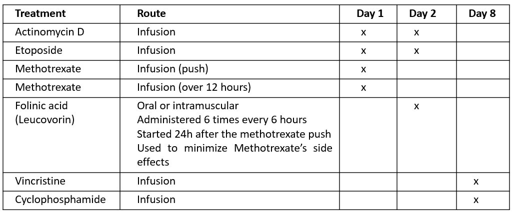
EMA-CO
 
  • This schedule consists of a combination of several medications:
     
 
  • Every treatment week is followed by a week without treatment
  • Treatment is repeated every 2 weeks until hCG levels normalize, followed by at least two mandatory consolidation cycles
  • You might be hospitalized for the administration of the therapy, depending on the guidelines of the center where you are treated 

Other treatment schedules might be proposed to you, based on your situation. If so, your treating physician will discuss these with you in more detail. 
 
2. SURGICAL TREATMENT
 
  • Second curettage: may be considered if residual trophoblastic tissue is seen on ultrasound; cure rate for GTN of approximately 40%.
     
  • Hysterectomy: surgical removal of the uterus; an alternative treatment in women who have completed childbearing and have disease confined to the uterus.
     
 Please feel free to contact us if you have any questions about your treatment.

The possible side effects depend on the type of treatment you receive. Not all patients experience side effects and their intensity can vary.

Some side effects that may occur with different treatments include:
 
  • Low white blood cell count (neutropenia), which can increase the risk of infections
  • Low platelet count (thrombocytopenia), which can affect blood clotting
  • Nausea and vomiting
  • Mouth sores – good oral hygiene is important; use a soft toothbrush and mild toothpaste and a prescribed mouthwash if needed
  • Fatigue
  • Liver or kidney effects – your doctor will advise you on which medications to avoid during treatment
  • Lung inflammation (pneumonitis) – contact your doctor immediately if you experience shortness of breath or breathing difficulties
  • Hair loss (alopecia) – this may occur with some treatments

Your treating physician will explain which side effects are most relevant to your treatment and how to reduce discomfort. Do not hesitate to contact your care team if you have any concerns.

If, during treatment, your hCG levels increase or stop decreasing without returning to normal, this means that the treatment is not having the desired effect. 

In this situation, a different treatment will be proposed. Your treating physician will explain the reasons for this change and discuss the new treatment plan with you in detail. 

Follow-up after GTN is different from follow-up after a molar pregnancy. 
 
  • Low-risk diseases 
In low-risk gestational trophoblastic neoplasia (GTN), once the hCG level has returned to normal, regular follow-up is recommended for at least one year, with monthly blood tests. 

Any remaining findings seen on imaging tests (such as ultrasound or scans) do not need further treatment as long as the hCG level remains normal. 

The risk of the disease coming back is low, but it can vary depending on the original diagnosis (the risk is slightly higher after choriocarcinoma) and on whether lung involvement was present at the time of diagnosis. 

If a recurrence does occur, it most often happens within the first year, which is why close follow-up during this period is important. Between year 2 and year 5, we propose hCG monitoring at least bi-yearly.

After a subsequent pregnancy, 6 weeks after the end of pregnancy, your hCG level should have normalized and a repeat hCG is recommended at this time to ensure no reactivation of GTN.
 
  • High-risk diseases
     
Protocols for hCG follow-up varied in frequency and duration between GTD centers. However, international consensus was reached to monitor the hCG at least monthly for a year or more (generally 18 months) after 4-8 weeks of normal hCG values after completion of treatment. 

If a recurrence does occur, it most often happens within the first year, which is why close follow-up during this period is important. Between year 2 and year 5, we propose hCG monitoring at least bi-yearly.

After a subsequent pregnancy, 6 weeks after the end of pregnancy, hCG should have normalized and a repeat hCG is recommended at this time to ensure no reactivation of GTN.

Please feel free to contact the responsible person in your reference center in case you need support during or after the treatment of a GTN. 

The contact persons for the Flemish reference center are:
Tine Op de beeck                   016/342 923           tine.opdebeeck@uzleuven.be
Ellen Reynders                         016/342 996           ellen.reynders@uzleuven.be
Joke De Roover                      016/347 419           joke.deroover@uzleuven.be

The contact persons for the French reference center are:
Frédéric Goffin                     info-fr@mole-chorio.befgoffin@chuliege.be
Sophie Schoenen                 info-fr@mole-chorio.bes.schoenen@chuliege.be
Christelle Leclercq               04/321.65.10 – 04/321.36.72

Please feel free to contact the responsible person in your reference center in case you need support after the diagnosis of a molar pregnancy or a GTN. 

The contact persons for the Flemish reference center are:
Tine Op de beeck                   016/342 923           tine.opdebeeck@uzleuven.be
Ellen Reynders                         016/342 996           ellen.reynders@uzleuven.be
Joke De Roover                      016/347 419           joke.deroover@uzleuven.be

The contact persons for the French reference center are:
Frédéric Goffin                     info-fr@mole-chorio.befgoffin@chuliege.be
Sophie Schoenen                 info-fr@mole-chorio.bes.schoenen@chuliege.be
Christelle Leclercq               04/321.65.10 – 04/321.36.72 

Follow-up


After conception, the fertilized egg is surrounded by trophoblastic cells, which attach it to the uterine wall and later form the placenta. These cells produce the hCG hormone (human chorionic gonadotropin), which is used to detect pregnancy. 

In a molar pregnancy or gestational trophoblastic neoplasia (GTN), these cells grow abnormally and produce excessive amounts of hCG. This hormone therefore serves as a key marker to monitor the course of the disease. 

After treatment or miscarriage, the hCG level should gradually return to normal, indicating that the molar tissue has disappeared. In about 80–90% of women, this happens spontaneously. However, if the hCG level plateaus, decreases too slowly or rises again, it may signal that the disease is persisting or progressing to a GTN, which requires specialized treatment (most often chemotherapy).

For this reason, regular blood tests are essential.
  • Partial mole 
You will need to have a blood test at your usual laboratory at least once every two weeks until hCG levels normalize (threshold varies depending on your laboratory). 
This normalization must be confirmed by at least one test within the month following normalization. 
This concludes the monitoring of a partial mole. In some cases, eg. when your hCG normalization took more than 8 weeks, a longer follow-up might be advised. 
 
  • Complete mole
A blood test at your usual laboratory is recommended at least once every two weeks until hCG levels normalize (threshold varies depending on your laboratory). 
After normalization, monthly tests will be performed for a duration of 6 months. 
 
  • Gestational trophoblastic neoplasia 
Follow-up after GTN is different from follow-up after a molar pregnancy.


Low-risk diseases 
In low-risk gestational trophoblastic neoplasia (GTN), once the hCG level has returned to normal, regular follow-up is recommended for at least one year, with monthly blood tests. 
Any remaining findings seen on imaging tests (such as ultrasound or scans) do not need further treatment as long as the hCG level remains normal. 
The risk of the disease coming back is low, but it can vary depending on the original diagnosis (the risk is slightly higher after choriocarcinoma) and on whether lung involvement was present at the time of diagnosis. 
If a recurrence does occur, it most often happens within the first year, which is why close follow-up during this period is important. Between year 2 and year 5, we propose hCG monitoring at least bi-yearly.

After a subsequent pregnancy, 6 weeks after the end of pregnancy, hCG should have normalized and a repeat hCG is recommended at this time to ensure no reactivation of GTN occurred. 

High-risk diseases 
Protocols for hCG follow-up varied in frequency and duration between GTD centers. However, international consensus was reached to monitor the hCG at least monthly for a year or more (generally 18 months) after 4-8 weeks of normal hCG values and completion of treatment. 
If a recurrence does occur, it most often happens within the first year, which is why close follow-up during this period is important. Between year 2 and year 5, we propose hCG monitoring at least bi-yearly.

After a subsequent pregnancy, 6 weeks after the end of pregnancy, hCG should have normalized and a repeat hCG is recommended at this time to ensure no reactivation of GTN occurred.

In a normal pregnancy, the hormone hCG (human chorionic gonadotropin) is mainly present in its intact “beta” form, which is the one detected by standard pregnancy tests

In molar pregnancies or gestational trophoblastic diseases (GTN), however, the body produces several different forms of hCG — including fragmented or less glycosylated variants. Because standard tests are designed to detect only the usual form seen in normal pregnancies, they can sometimes give false-negative results in these diseases. 

To ensure reliable monitoring, we therefore measure total hCG, which includes all molecular forms of the hormone. It is also very important that all your blood samples are analyzed in the same laboratory, using the same test method and detection threshold, so that the results can be accurately compared over time. 

In certain situations, you may also be asked to provide a urine sample for hCG testing. This can help confirm the results if we suspect that antibodies in your blood are interfering with the test, since such antibodies are not present in urine. 

A recurrence of gestational trophoblastic neoplasia (GTN), or progression from a molar pregnancy to GTN, can occur several months after the initial diagnosis. For this reason, it is essential to monitor hCG levels over time to make sure they remain normal. 
 
Partial moles:
  • Once the hCG level has returned to normal, this result must be confirmed by a second blood test within one month.
  • If the second test is also normal, usually no further follow-up is required.
  • This usually marks the end of follow-up for a partial mole.
  • In some cases, eg. when your hCG normalization took more than 8 weeks, a longer follow-up might be advised.
  • After a subsequent pregnancy6 weeks after the end of pregnancy, your hCG should have normalized and a repeat hCG is recommended at this time to ensure no reactivation of the GTN occurred. 

Complete moles:
  • Because the risk of developing GTN is higher after a complete mole, monthly blood tests to measure hCG are recommended for 6 months after normalization.
  • If hCG remains normal throughout this 6-month period, follow-up can be stopped.
  • Timing of follow-up:
    • If your hCG normalizes within 8 weeks after the first curettage, the follow-up lasts 6 months from the date of the first curettage.
    • If your hCG normalizes more than 8 weeks after the first curettage, the follow-up lasts 6 months from the date of hCG normalization.
  • After a subsequent pregnancy6 weeks after the end of pregnancy, your hCG should have normalized and a repeat hCG is recommended at this time to ensure no reactivation of the GTN occurred.
 
Gestational trophoblastic neoplasia:
  • At least 12 months for low-risk diseases and 18 months for high-risk diseases. After a subsequent pregnancy, 6 weeks after the end of pregnancy, your hCG should have normalized and a repeat hCG is recommended at this time to ensure no reactivation of the GTN occurred. 

You can find more information on the hCG testing frequency in the section ‘How often do I need to have my hCG levels tested?’

You will need to use contraception to avoid becoming pregnant from the time of the curettage until the end of the follow-up period. This is important to allow reliable monitoring of your hCG levels. 

In most cases, you will be prescribed an oral contraceptive pill. 

If you have a medical contraindication to oral contraceptives, or if you prefer a different method of contraception, your doctor will discuss safe and effective alternative options with you. 

An intrauterine contraceptive device (IUCD) can be inserted only if and when your doctor considers it appropriate.

We monitor your blood hCG levels to track your molar pregnancy or GTN. Even after hCG levels return to normal, there is a small risk of recurrence, mainly in the first months. Close monitoring during this period is therefore essential. As a pregnancy also increases hCG levels, you should avoid becoming pregnant during follow-up, as this could delay the detection and treatment of a recurrence. 

More information on the testing and follow-up schedule can be found in the section ‘How often do I need to have my hCG levels tested?’.

A new pregnancy is usually permitted as soon as hCG normalization is confirmed after a partial mole. In some cases, eg. when your hCG normalization took more than 8 weeks, a longer follow-up, and thus a longer delay of a next pregnancy, might be advised

For complete moles, a waiting period of 6 months is required, during which hCG levels will be monitored monthly. 

For gestational trophoblastic neoplasia, a waiting period of 12 to 18 months is generally recommended after hCG normalization and completion of chemotherapy. However, each case requires expert evaluation for optimal management. 

More information on the testing and follow-up schedule can be found in the section ‘How often do I need to have my hCG levels tested?’.

Please feel free to contact the responsible person in your reference center in case you need support after the diagnosis of a molar pregnancy or a GTN. 

The contact persons for the Flemish reference center are:
Tine Op de beeck                   016/342 923           tine.opdebeeck@uzleuven.be
Ellen Reynders                         016/342 996           ellen.reynders@uzleuven.be
Joke De Roover                      016/347 419           joke.deroover@uzleuven.be

The contact persons for the French reference center are:
Frédéric Goffin                     info-fr@mole-chorio.befgoffin@chuliege.be
Sophie Schoenen                 info-fr@mole-chorio.bes.schoenen@chuliege.be
Christelle Leclercq               04/321.65.10 – 04/321.36.72 

Please feel free to contact the responsible person in your reference center in case you need support after the diagnosis of a molar pregnancy or a GTN. 

The contact persons for the Flemish reference center are:
Tine Op de beeck                   016/342 923           tine.opdebeeck@uzleuven.be
Ellen Reynders                         016/342 996           ellen.reynders@uzleuven.be
Joke De Roover                      016/347 419           joke.deroover@uzleuven.be

The contact persons for the French reference center are:
Frédéric Goffin                     info-fr@mole-chorio.befgoffin@chuliege.be
Sophie Schoenen                 info-fr@mole-chorio.be   / s.schoenen@chuliege.be
Christelle Leclercq               04/321.65.10 – 04/321.36.72 

Future implications


We strongly advise you to wait before trying to become pregnant until your hCG levels have returned to normal and you have completed the recommended follow-up period. This allows any possible recurrence of the disease to be detected as early as possible. 

A new pregnancy is usually permitted as soon as hCG normalization is confirmed after a partial mole. In some cases, eg. when your hCG normalization took more than 8 weeks, a longer follow-up, and thus a longer delay of a next pregnancy, might be advised

For complete moles, a waiting period of 6 months is required, during which hCG levels will be monitored monthly. 

For gestational trophoblastic neoplasia, a waiting period of 12 to 18 months is generally recommended after hCG normalization and completion of chemotherapy. However, each case requires expert evaluation for optimal management. 

More information on the testing and follow-up schedule can be found in the section ‘How often do I need to have my hCG levels tested?’.

The chemotherapy treatments used do not affect fertility and do not increase the risk of congenital abnormalities. However, we recommend waiting one full year before starting a new pregnancy. This allows your body to fully eliminate the treatment and recover, so that a future pregnancy can take place under the best possible conditions

Some side effects may persist long term or, in rare cases, be permanent. This is mainly the case for neuropathy, a nerve-related side effect that can occur with paclitaxel, one of the chemotherapy drugs sometimes used in combination treatments. Neuropathy usually causes reduced sensation, tingling or cramping in the fingers, toes or soles of the feet. If this occurs during treatment, symptoms usually improve gradually in the months after treatment ends, but they may not completely disappear. 

If you received paclitaxel, etoposide, or actinomycin, you may have experienced hair loss (alopecia). When your hair grows back, its texture may change. For example, straight hair may become curly, or vice versa. These changes can sometimes be permanent. 

Please do not hesitate to contact us in case you have questions regarding any side effects that remain present.

The risk of having another molar pregnancy or GTN is about 1%, which is 10 to 20 times higher than in the general population. If you have had two previous molar pregnancies, the risk of having a third increases to 15–20%

If you are diagnosed with a second molar pregnancy, genetic testing may be offered to determine whether an underlying cause can be identified.

There is no international consensus on this topic. However, we recommend an early ultrasound examination in any future pregnancy to confirm the presence of a normal, viable fetus. 

If no viable fetus is identified, we recommend a pathological examination of the curettage tissue.

Only a very small percentage of women have a familial predisposition for recurrent molar pregnancies. This rare condition is known as familial recurrent hydatidiform mole (FRHM) and due to genetic mutations (NLRP7, KHDC3L). 

It is important to inform your doctor if any of your relatives have experienced a molar pregnancy or GTN. This allows genetic counselling to be offered and, if needed, further advice regarding future pregnancies.

Please feel free to contact the responsible person in your reference center in case you need support during or after the treatment of a GTN. 

The contact persons for the Flemish reference center are:
Tine Op de beeck                   016/342 923           tine.opdebeeck@uzleuven.be
Ellen Reynders                         016/342 996           ellen.reynders@uzleuven.be
Joke De Roover                      016/347 419           joke.deroover@uzleuven.be

The contact persons for the French reference center are:
Frédéric Goffin                     mole.chorio@chuliege.befgoffin@chuliege.be
Sophie Schoenen                 mole.chorio@chuliege.bes.schoenen@chuliege.be
Christelle Leclercq               04/321.65.10 – 04/321.36.72 

Please feel free to contact the responsible person in your reference center in case you need support after the diagnosis of a molar pregnancy or a GTN. 

The contact persons for the Flemish reference center are:
Tine Op de beeck                   016/342 923           tine.opdebeeck@uzleuven.be
Ellen Reynders                         016/342 996           ellen.reynders@uzleuven.be
Joke De Roover                      016/347 419           joke.deroover@uzleuven.be

The contact persons for the French reference center are:
Frédéric Goffin                     info-fr@mole-chorio.befgoffin@chuliege.be
Sophie Schoenen                 info-fr@mole-chorio.bes.schoenen@chuliege.be
Christelle Leclercq               04/321.65.10 – 04/321.36.72